Atomic force microscopy (AFM) offers a method for
label-free imaging of nanoscale biomolecular dynamics to solve
biological questions that cannot be addressed via other bioimaging
methods including fluorescence and scanning electron microscopy.
Since such imaging methods are only possible for biological systems
extracted from cells or reconstructed on solid substrates,
nanodynamics within living cells largely remain inaccessible with
existing bioimaging methods. In a new report now published in
Science Advances, Marcos Penedo and a research team in Nanolife
Science and biotechnology at the Kanazawa University in Japan,
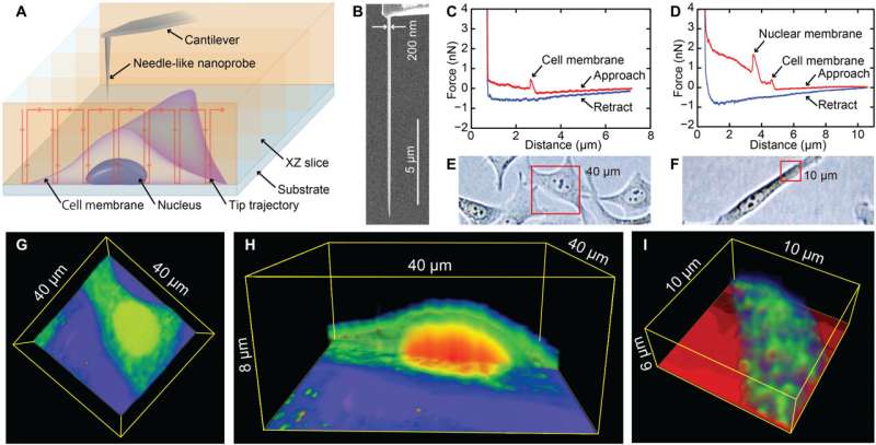
overcame the limits of bioimaging by using nanoendoscopy-AFM.
During the process, they inserted a needle-like probe into a living
cell to present actin fiber, three-dimensional (3D) maps and 2D
nanodynamics of the inner scaffold of the membrane with
undetectable changes in cell viability. Unlike earlier AFM methods,
the nanoprobe directly accessed the target intracellular components
and explored the capabilities of AFM, including high-resolution
imaging, nanomechanical mapping and molecular recognition to expand
the observable range of intracellular structures in living
cells.
Visualizing intracellular nanostructures of living cells
using nanoendoscopy-AFM