Raman spectroscopy is widely used in analytical
sciences to identify molecules via their structural fingerprint. In
the biological context the Raman response provides a valuable
label-free specific contrast that allows distinguishing different
cellular and tissue contents. Unfortunately, spontaneous Raman
scattering is very weak, over ten orders of magnitude weaker than
fluorescence. Unsurprisingly, fluorescence microscopy is often the
preferred choice for applications such as live cell imaging.
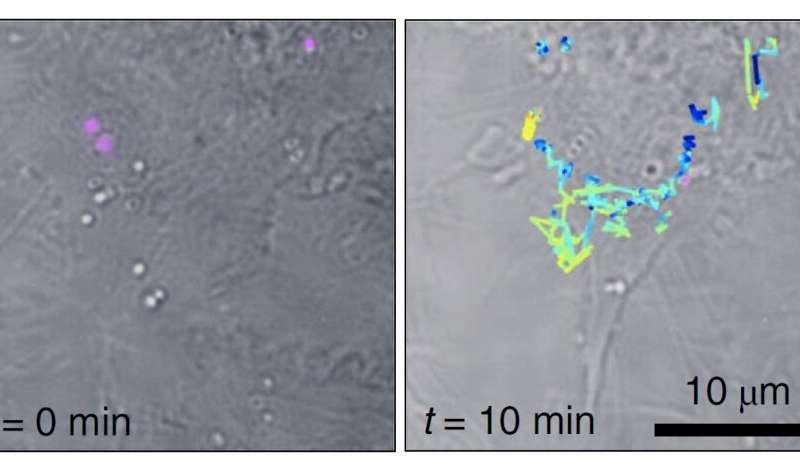
Luckily, Raman can be enhanced dramatically on metal surfaces or in
metallic nanogaps and this surface enhanced Raman scattering (SERS)
can even overcome the fluorescence response. Nanometric SERS probes
are thus promising candidates for biological sensing applications,
preserving the intrinsic molecular specificity. Still, the
effectiveness of SERS probes depends critically on the particle
size, stability and brightness, and, so far, SERS-probe based
imaging is rarely applied.